NeuroCovid: schade en protectie
21 May, 2021
Door: Fokje Russchen
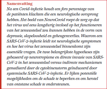
 Al vrij snel werd in de Covid-19-pandemie melding gemaakt van neurologische verschijnselen en kwam de term NeuroCovid in gebruik. Het beeld van NeuroCovid roept de zorg op dat het virus langdurig invloed zou kunnen hebben op het functioneren van het zenuwstelsel. En wel in de vorm van depressie, slapeloosheid en geheugenverlies. Het zou zelfs een rol kunnen spelen in het ontstaan van neurodegeneratieve aandoeningen zoals parkinson, narcolepsie en multiple sclerose.1
Er zijn meerdere epidemische virussen bekend die zenuwweefsel kunnen infecteren, bijvoorbeeld polio. Ook de influenza-pandemie van 1918, de SARS-uitbraak van 2003 en de MERS-uitbraak van 2012 – de laatste twee veroorzaakt door coronavirussen – leidden tot neurologische klachten die voor een deel pas maanden tot jaren later naar voren kwamen. De neurologische klachten van Covid-19 variëren van mild tot ernstig; van hoofdpijn, gestoorde reuk en/of smaak, myalgie en vermoeidheid of slaperigheid tot encefalopathie, ischemische of hemorragische beroerte, epilepsie, hypoxisch-ischemische hersenschade, het syndroom van Guillain-Barré en andere auto-immuun syndromen. In een retrospectief onderzoek in Wuhan werden neurologische symptomen gezien bij 36,4% van het totaal aantal opgenomen Covid-19-patiënten.2 In een onlangs gepubliceerde analyse over ruim 200.000 cases uit de VS bleek 34% van de Covid-19-patiënten neurologische of psychiatrische restverschijnselen te hebben (zie tabel 1).3
Het is duidelijk dat het virus voornamelijk de ACE 2-receptor gebruikt om menselijke cellen binnen te dringen. Covid-19 is vooral een ziekte van de luchtwegen en ACE 2 komt hier dan ook in grote hoeveelheden voor. Het zit daarnaast op cellen in vele andere organen, zo ook in hersenweefsel.4 Met name in cardiorespiratoire centra in de hersenstam.5 De resultaten van onderzoeken naar de vraag of ACE 2-receptoren op zenuwcellen voorkomen spreken elkaar echter tegen.6,7 SARS-CoV-2-deeltjes zijn post mortem in de hersenen gevonden in microvaten, de plexus choreoïdus, de hersenvliezen en, door sommigen, in neuronen.
Er zijn twee hypotheses die verklaren hoe het virus het zenuwweefsel binnentreedt. De eerste gaat uit van neurotropisme en directe invasie van SARS-CoV-2 in het zenuwstelsel: de neurogene route. De tweede gaat uit van indirecte effecten door de cytokinenstorm, geïnduceerd door de systemische infectie: de hematogene route.
Lees het gehele artikel vanaf pagina 22 in OrthoFyto 3/21.
Wilt u het gehele artikel als PDF bestand ontvangen? Bestel het dan hier voor € 3,50.
Bronvermelding:
1.Schirinzi T,Landi D, Liguori C. COVID-19: dealing with a potential risk factor for chronic neurological disorders. J Neurol. 2020 Aug 27 : 1–8.
2.Mao L., Jin H., Wang M., Hu Y., Chen S., He Q., Chang J., Hong C., Zhou Y., Wang D., et al. Neurologic Manifestations of Hospitalized Patients With Coronavirus Disease 2019 in Wuhan, China. JAMA Neurol. 2020.
3. Taquet, M, Geddes J, Husain,M Luciano,S Harrison P. Six-month Neurological and Psychiatric Outcomes in 236,379 Survivors of COVID-19. MedRXiv Lancet Psychiatry 6 april 2021.
4. Kehoe G, Wong S, Al Mulhim N, Palmer LE, Miners JS. Angiotensin-converting enzyme 2 is reduced in Alzheimer's disease in association with increasing amyloid-beta and tau pathology. Alzheimer Res Ther. 2016;8(1):50.
5. Xia H., Lazartigues E. (2008). Angiotensin-converting enzyme 2 in the brain: properties and future directions. J. Neurochem. 107, 1482–1494.
6. Xu J, Lazartigues E. Expression of ACE2 in Human Neurons Supports the Neuro-Invasive Potential of COVID-19 Virus. Cell Mol Neurobiol. 2020 Jul 4 : 1–5.
7. Song E, et al. 2021. Neuroinvasion of SARS-CoV-2 in human and mouse brain. J Exp Med 1 March 2021; 218 (3): e20202135.
8. Klopfenstein T., Kadiane-Oussou N.J., Toko L., Royer P.Y., Lepiller Q., Gendrin V., Zayet S. Features of anosmia in COVID-19. Med. Mal. Infect. 2020;50:436–439.
9. Brann, D. H. et al. Non-neuronal expression of SARS-CoV-2 entry genes in the olfactory system suggests mechanisms underlying COVID-19-associated anosmia. Sci. Adv. 6, eabc5801 (2020).
10. Meinhardt J, Radke J, Dittmayer C, Franz J, Thomas C, Mothes, R. Laue M, Schneider J.et al. Olfactory transmucosal SARS-CoV-2 invasion as a port of central nervous system entry in individuals with COVID-19. Nat. Neurosci. 2020, 1–8.
11. Briguglio M, et al. (2020). Disentangling the Hypothesis of Host Dysosmia and SARS-CoV-2: The Bait Symptom That Hides Neglected Neurophysiological Routes. June 2020, Frontiers in Physiology 11:671.
12. Helms J., Kremer S., Merdji H., Clere-Jehl R., Schenck M., Kummerlen C., Collange O., Boulay C., Fafi-Kremer S., Ohana M., et al. Neurologic Features in Severe SARS-CoV-2 Infection. N. Engl. J. Med. 2020;382:2268–2270.
13. Kanberg, N. et al. Neurochemical evidence of astrocytic and neuronal injury commonly found in COVID-19. Neurology 95, e1754–e9 (2020).
14. Pilotto, A., Padovani, A. & ENCOVID-BIO Network. Reply to the letter ‘COVID-19-associated encephalopathy and cytokine-mediated neuroinflammation’. Ann. Neurol. 88, 861–862 (2020).
15. Murta V, Villarreal A, Ramos A. Severe Acute Respiratory Syndrome Coronavirus 2 Impact on the Central Nervous System: Are Astrocytes and Microglia Main Players or Merely Bystanders? ASN Neuro. 2020 Jan-Dec; 12.
16. Reynolds J.Mahajan H. SARS-COV2 Alters Blood Brain Barrier Integrity Contributing to Neuro-Inflammation. J Neuroimmune Pharmacol. 2021 Jan 6 : 1–3.
17. Wang et al. 2021. ApoE-Isoform-Dependent SARS-CoV-2 Neurotropism and Cellular Response Cell Stem. Cell. 2021 Feb 4; 28(2): 331–342.e5.Pub.
18. Lippi A, Domingues R, Setz C, Outeiro TF, Krisko A. (2020). SARS-cov-2: at the crossroad between aging and neurodegeneration. Mov Disord 35, 716–720.
19. Fazzini E, Fleming J, Fahn S. (1992). Cerebrospinal fluid antibodies to coronavirus in patients with Parkinson’s disease. Mov Disord 7, 153–158.
20. Vries R. (2021) Intranasal fusion inhibitory lipopeptide prevents direct-contact SARS-CoV-2 transmission in ferrets. Science 17 Feb 2021.
21. Rezaeiyan D, Darou Laboratories Company. Evaluation efficacy of a herbal combination (contains Glycyrrhiza glabra, Mentha pulegium and Urtica) in controlling symptoms in patients with COVID-19. Iranian Registry of Clinical Trials.
22. Ellul, M. A. et al. Neurological associations of COVID-19. Lancet Neurol. 19, 767–83 (2020).
23. Brann, D. H. et al. Non-neuronal expression of SARS-CoV-2 entry genes in the olfactory system suggests mechanisms underlying COVID-19-associated anosmia. Sci. Adv. 6, eabc5801 (2020).
24. Liang Chen et al. (2020). A Novel Combination of Vitamin C, Curcumin and Glycyrrhizic Acid Potentially Regulates Immune and Inflammatory Response Associated with Coronavirus Infections: A Perspective from System Biology. Analysis Nutrients. 2020 Apr; 12(4): 1193.Published online 2020 Apr 24.
25. Gao Y, Fu R, Wang J, Yang X, Wen L, Feng J. Resveratrol mitigates the oxidative stress mediated by hypoxic-ischemic brain injury in neonatal rats via Nrf2/HO-1 pathway. Pharm Biol. 2018;56:440–9.
26. Liu WC, Wang X, Zhang X, Chen X, Jin X. Melatonin supplementation, a strategy to prevent neurological diseases through maintaining integrity of blood brain barrier in old people. Front Aging Neurosci. 2017;9:165.
27. Prapimpun Wongchitrat, Mayuri Shukla, Ramaswamy Sharma, Piyarat Govitrapong, Russel J. Reiter. Role of Melatonin on Virus-Induced Neuropathogenesis—A Concomitant Therapeutic Strategy to Understand SARS-CoV-2 Infection Antioxidants (Basel). 2021 Jan; 10(1): 47. Published online 2021 Jan 2.
28. Fakhri S, Nouri Z, Moradi S, Farzaei M. Astaxanthin, COVID‐19 and immune response: Focus on oxidative stress, apoptosis and autophagy. Phytother Res. 2020 Aug 4 : 10.1002/ptr.6797.
29. Cengiz, M., Borku Uysal, B., Ikitimur, H., Ozcan, E., Islamoğlu, M. S., Aktepe, E., Yavuzer, H., & Yavuzer, S. (2020). Effect of oral L-Glutamine supplementation on Covid-19 treatment. Clinical Nutrition Experimental, Volume 33, October 2020, Pages 24-31.
30. Name et al. Vitamin D, zinc and glutamine: Synergistic action with OncoTherad immunomodulator in interferon signaling and COVID 19 (Review). Int J Mol Med . 2021 Mar;47(3).
